
In clinical laboratories and cytopathology, specimen preparation plays an important role in cell morphology assessment and subsequent microscopic analysis. For body fluid specimens such as cerebrospinal fluid (CSF), pleural effusions, ascites, and urine, effective cell recovery, minimal specimen loss, and consistent cell distribution are essential to producing high-quality cytology slides.
These requirements are particularly important for low-cellularity specimens, such as CSF, in which the number of available cells may be limited and some cells can be vulnerable to morphological changes during processing. Manual smear preparation and conventional centrifugation followed by separate slide preparation may introduce variability, potentially resulting in uneven cell distribution, overlapping cells, or cell loss.
A cytocentrifuge integrates centrifugal cell deposition with slide preparation. With programmable controls, stable centrifugal operation, and dedicated slide preparation components, it helps laboratories standardize specimen processing and prepare slides suitable for subsequent staining and microscopic examination.
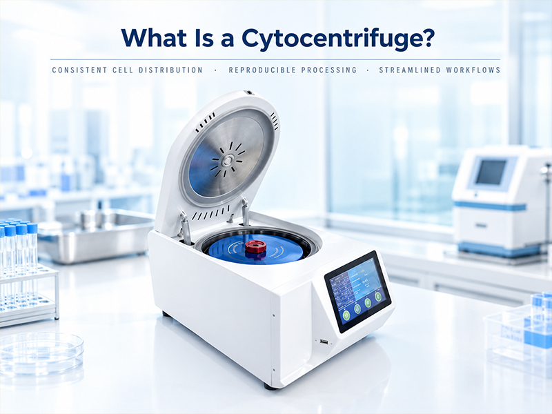
What Is a Cytocentrifuge? Working Principle and Key Benefits
A cytocentrifuge is a specialized laboratory instrument used to concentrate cells from liquid specimens onto a designated area of a microscope slide. By applying controlled centrifugal force, suspended cells are deposited onto the slide, creating a preparation suitable for cytological staining and microscopic evaluation.
Unlike manual smear techniques, which depend heavily on the operator's technique, a cytocentrifuge allows users to control parameters such as rotational speed, centrifugation time, and relative centrifugal force (RCF). This helps reduce operator-dependent variation and supports more reproducible specimen preparation.
Key benefits include:
● More consistent cell distribution: Appropriate centrifugation conditions and slide preparation components help reduce uneven deposition and localized cell accumulation.
● Improved cell recovery: Centrifugal deposition helps collect suspended cells onto the designated slide area while reducing cell loss associated with certain specimen transfer steps.
● Reproducible processing: Programmable settings can be stored and reused, helping laboratories maintain consistency across operators and batches.
● Streamlined laboratory workflows: Automated operation reduces repetitive manual handling and supports routine cytology specimen processing.
● Enhanced specimen handling safety: Sealed rotors, lid-locking mechanisms, and appropriate cleaning and disinfection procedures help reduce the risk of specimen leakage and biological exposure.
Actual preparation quality depends on specimen characteristics, cell concentration, pretreatment methods, centrifugation parameters, and slide preparation components. These factors should be evaluated for each intended application.
Why Does CSF Cytology Require Careful Specimen Preparation?
Cerebrospinal fluid (CSF) is a representative application for cytocentrifugation because it typically contains relatively few cells. In some clinical specimens, diagnostically relevant abnormal cells may also be present in limited numbers.
Consequently, CSF cytology requires careful attention to cell recovery, preservation of cellular morphology, and the distribution of cells on the prepared slide.
● Low cell counts make cell recovery particularly important.
Because CSF may contain only a limited number of cells, cell loss during specimen transfer, residual material left in containers, or slide preparation can affect the number of cells available for examination.
A cytocentrifuge helps concentrate suspended cells onto a designated slide area and can reduce the need for certain manual transfer steps. This supports a more consistent cell concentration workflow, although actual recovery rates depend on the specimen, rotor design, preparation components, and operating conditions.
● Centrifugation conditions must balance cell recovery and morphological preservation.
Insufficient centrifugal force may result in incomplete cell sedimentation, whereas excessive force or inappropriate processing time may affect the morphology of some cells.
For this reason, CSF preparation requires suitable centrifugation conditions rather than simply maximizing rotational speed or processing time. Programmable control helps laboratories establish and repeat validated protocols, reducing variability in routine operation.
● Cell distribution affects microscopic examination.
When cells accumulate in localized areas, overlapping cells may make morphological assessment more difficult. Conversely, sparse or uneven deposition can increase the area that needs to be examined.
Appropriate centrifugation conditions, combined with suitable slide preparation components, help promote more consistent cell distribution and provide a better starting point for subsequent staining and microscopic examination.
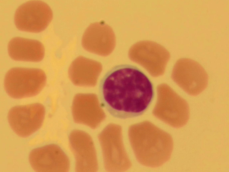
How a Cytocentrifuge Improves CSF Slide Preparation
Cerebrospinal fluid (CSF) often contains a relatively low number of cells, making efficient cell concentration and careful slide preparation important for cytological examination. A cytocentrifuge streamlines this process by concentrating cells onto a defined area of a microscope slide, helping laboratories reduce manual handling steps and achieve more consistent specimen preparation.
● Improving Cell Recovery and Reducing Specimen Loss
When preparing CSF slides, cell loss can occur during specimen transfer, concentration, and other manual handling steps. These losses may be particularly significant when the original specimen contains very few cells.
A cytocentrifuge uses controlled centrifugal force to deposit cells from a liquid specimen onto a designated area of the slide. By combining cell concentration and slide preparation in a single workflow, it can reduce the need for additional transfer steps and help minimize handling-related specimen loss.
Actual cell recovery depends on factors such as specimen characteristics, pretreatment procedures, centrifugation settings, and the compatibility of the slide preparation components. Recovery performance should therefore be evaluated using the intended specimen types and validated laboratory procedures.
● Controlling Centrifugation Parameters for Consistent Preparation
Appropriate centrifugation settings help balance effective cell deposition with the preservation of cellular morphology. The optimal sample volume, rotational speed, and centrifugation time depend on the cellularity of the specimen and its pretreatment history.
According to the Welso WTP-2 cytocentrifuge manual, CSF samples that have undergone cell counting and concentration can use 15–20 μL of sample at 1,200 rpm for 3 minutes as reference settings. For samples that have not undergone these pretreatment steps, the sample volume may be increased to approximately 30–50 μL, depending on specimen characteristics and the observed slide preparation results.
These settings are reference values rather than universal requirements. Laboratories should evaluate the resulting slides and adjust operating parameters according to their specimen types, rotor configuration, and validated procedures. Since rpm and relative centrifugal force (RCF, expressed as ×g) are not interchangeable, the effective centrifugal force also depends on the rotor radius.
With programmable centrifugation settings, the WTP-2 helps laboratories establish and repeat suitable operating conditions, supporting a more standardized preparation workflow and reducing variation caused by inconsistent manual operation.
● Promoting More Uniform Cell Deposition
A cytocentrifuge directs cells toward a defined deposition area on the microscope slide, helping produce a concentrated cellular region for subsequent staining and microscopic examination. When the sample volume, centrifugation conditions, and slide preparation components are appropriately matched, the resulting cell distribution may be more suitable for routine cytological assessment than preparations relying solely on manual smearing.
The quality of cell deposition still depends on specimen composition, cell characteristics, and the preparation method. Appropriate staining, specimen handling, and laboratory quality control remain essential for obtaining interpretable slides.
● Supporting a More Standardized CSF Preparation Workflow
By integrating cell concentration and slide preparation, a cytocentrifuge can simplify routine CSF processing, reduce certain manual handling steps, and help laboratories reproduce established preparation conditions. These advantages are particularly relevant when working with specimens that contain limited numbers of cells.
For reliable results, centrifugation parameters should be selected according to the specimen and preparation method rather than applied uniformly to every sample. Used alongside validated laboratory procedures, the Welso WTP-2 provides a practical tool for supporting consistent CSF cytology slide preparation.
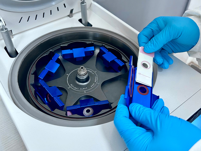
Key Features of the Welso Cytocentrifuge
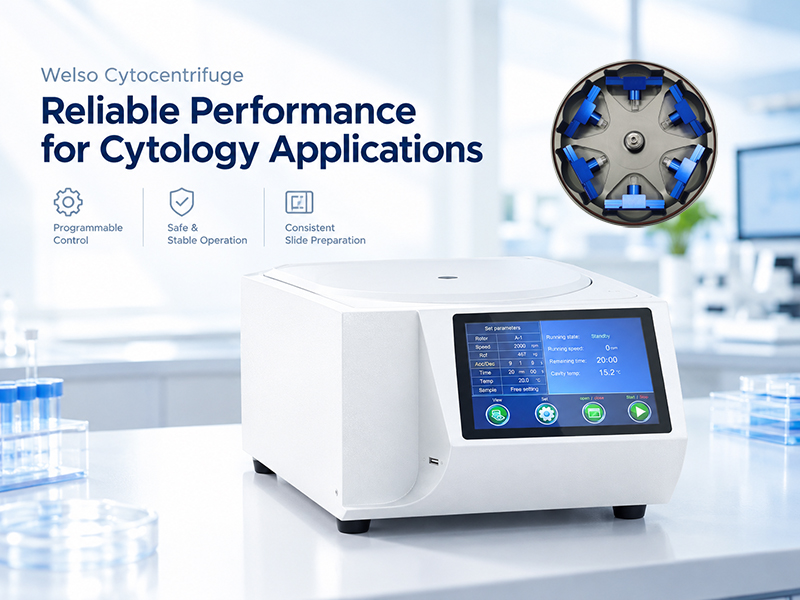
Designed for laboratory workflows that require consistent operation, convenient control, and appropriate specimen handling, the Welso cytocentrifuge incorporates programmable controls, a brushless drive system, and a sealed rotor design.
● Programmable Control and RCF Settings
The instrument uses microprocessor-based control and supports program storage and recall. Centrifugation parameters can be configured according to the requirements of the intended application.
Gradient centrifugation and RCF calculation and setting functions support the development of application-specific protocols, helping laboratories reduce repetitive setup and maintain consistent processing conditions.
● Brushless DC Motor for Stable Operation
The brushless DC motor supports smooth operation and low-noise performance without the routine carbon-brush maintenance required by brushed motors. It also helps avoid dust generated by carbon-brush wear.
● Stainless Steel Chamber for Easier Maintenance
The centrifuge chamber and designated components exposed to specimen-related vapors are made of 304 stainless steel, supporting routine cleaning and maintenance.
● Independent Exhaust System
An independent exhaust duct helps remove air and relevant vapors generated during operation. Where applicable, compatible filtration components can provide additional control of airborne contaminants.
Actual filtration performance and protective capabilities depend on the installed configuration and validated operating conditions.
● Seven-Inch Touchscreen for Convenient Operation
A 7-inch touchscreen provides an intuitive interface for parameter setting and operating status monitoring. One-touch automatic lid opening and closing helps simplify routine operation.
● Intelligent Lid Locking and Imbalance Protection
The intelligent lid-locking system uses a stepper motor to provide automatic engagement, combining electronic locking with a mechanical locking structure for enhanced operating safety.
Dynamic imbalance protection detects abnormal imbalance and initiates a stop according to the instrument's safety control logic, helping reduce the risks associated with excessive vibration and unstable operation.
● Precision-Machined Rotor and Sealed Sample Handling
The rotor and sample holders are precision-machined from aluminum alloy and treated with an anodized surface finish to improve wear and corrosion resistance.
The replaceable sealed rotor design allows different configurations to be used according to application requirements. Rotor installation and removal can be performed within a safety enclosure. A transparent rotor cover allows sample status to be viewed where the operating conditions permit.
Rotor components that are compatible with autoclaving may be sterilized according to the manufacturer's instructions and specified temperature, pressure, and cycle requirements.
The sealed design helps reduce the risk of specimen leakage and biological exposure. It should be used alongside appropriate personal protective equipment, cleaning, disinfection, and laboratory biosafety procedures.
● Easy-to-Clean Chamber and Fault Monitoring
The internal chamber is designed for convenient cleaning and maintenance. Safety alarms and fault codes help operators identify abnormal conditions and support routine troubleshooting.
Absorbent pads undergo dust-reduction processing, ultraviolet/ozone treatment, and vacuum packaging to help reduce the risk of introducing contaminants during use. Consumables should be stored, replaced, and handled according to the manufacturer's instructions.
Together, these features support convenient operation, routine maintenance, and controlled specimen processing. Specific performance characteristics, configurations, and applicable compliance standards should be confirmed against the technical documentation for the relevant model.
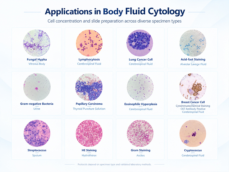
Applications in Body Fluid Cytology
Depending on the compatible rotor, slide preparation components, and validated protocols, the cytocentrifuge can be used to concentrate cells from various liquid specimens for cytological preparation.
Typical application areas include:
● Cerebrospinal fluid (CSF): Cell concentration and cytology slide preparation for low-cellularity specimens.
● Pleural, peritoneal, and pericardial effusions: Cell concentration and morphological examination of serous fluid specimens.
● Urine: Specimen preparation for urine cytology applications.
● Synovial fluid: Cell collection and slide preparation for joint fluid specimens.
● Sputum and bronchial specimens: Cytological preparation of respiratory specimens, with pretreatment where necessary to address viscosity and debris.
● Puncture aspirates and other suitable aspirated fluids: Cell concentration and slide preparation according to specimen requirements.
● Cervical cell specimens: Applicable thin-layer cytology workflows when compatible preparation components and validated methods are available.
Different specimen types vary in cellular composition, viscosity, debris content, and pretreatment requirements. The appropriate workflow should therefore be established according to the specimen, instrument configuration, and laboratory protocol rather than applying a single set of parameters to every sample.
Supporting Reliable Cytology Through Consistent Sample Preparation
High-quality cytology preparation requires more than collecting cells from a liquid specimen. It also involves preserving cellular morphology, promoting suitable cell distribution, maintaining consistent operating conditions, and managing biological risks.
The Welso cytocentrifuge combines programmable control, stable centrifugal operation, sealed rotor design, and multiple safety features to support automated preparation of CSF and other body fluid specimens.
For CSF cytology, appropriate centrifugation conditions and a validated preparation workflow can help improve cell recovery and distribution, providing a more suitable foundation for staining, microscopic examination, and cellular morphology assessment.
Welso provides laboratory sample preparation solutions designed to support reliable operation, convenient workflows, and appropriate specimen handling—helping laboratories improve process consistency and day-to-day efficiency.



Email: info@welsobio.com
Tel: +86 21 51096910
Head Office: No. 439 Jinglian Road, Minhang District, Shanghai,China, 201108
Branch: 1-1009, National Industrial Design Park, No. 599 Jianzhu Road, Binhu, Wuxi , Jiangsu, China, 214062

Follow official account